Several authors have previously reviewed the use of spectroscopy techniques for the optical characterization of diverse samples [1] [2]. Optical characterization techniques are defined as methods to study the form in which the electromagnetic radiation coming from a light source, interacts with a surface. Especially important is the range between UV light to the near Infrared Light (NIR) for the analysis of solid materials, particularly semiconductors.
Additionally, attention is paid to spectroscopy techniques, which allow for the study of ultrafast dynamics as a quantitative analysis of materials by means of relating the intensity versus the wavelength of the light that is irradiated by the sample. In spectroscopy, different modalities can be described attending to the quantity that is being measured (absorption, transmission, reflectance, scattering…). In these types of measurements, the spatial resolution becomes an essential aspect due to the fact that the spectral characteristics of the sample are being analysed. Moreover, the spatial resolution is limited by diffraction. As a result, different strategies can be used to go beyond the diffraction limit in spectroscopic techniques.
One approach comprises the use of a pump-probe system, which allows for nonlinear optical imaging leading to the improvement of the contrast, sensitivity and chemical specificity of the measurements. These advantages make it suitable for different applications in biological and material sciences. This technique is the Pump-Probe spectroscopy [3].
Pump-probe spectroscopy theory
Pump-probe spectroscopy is a modality of ultrafast optical spectroscopy. The theory behind this technique explains the process by which photons emitted by an ultrafast pulsed laser lead to the excited-state dynamics of the atoms that form a sample of interest. Being the excited-state dynamics three main processes: excited absorption, stimulated emission or ground-state depletion.
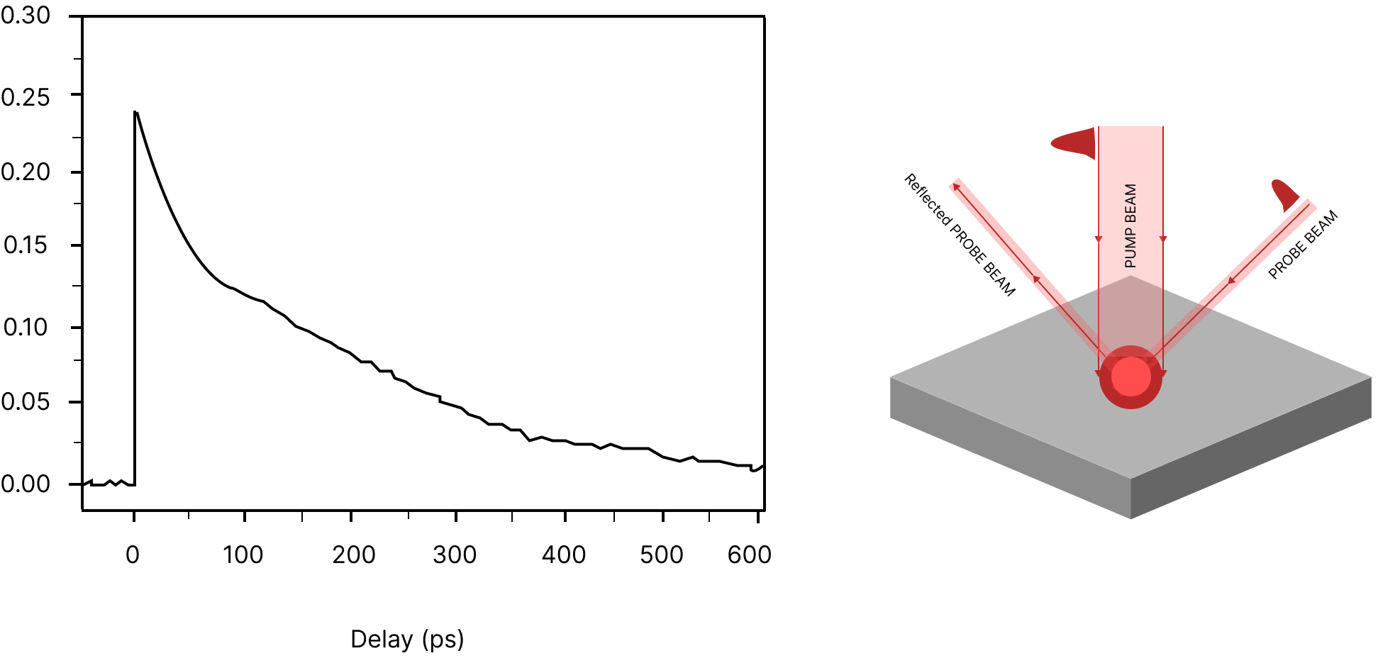
From a general point of view, the laser pulses are split into two beams: one stronger beam for the pumping of the atoms (pump beam), and a second beam for the monitoring of the changes in the optical properties (probe beam). Such optical properties are related to the reflection and transmission constants of the probe beam. Quantitative estimations of the changes in the optical properties are obtained by means of studying the time delay between the two beams (Figure 1).
What the Pump-probe technique brings into the table is that it is able to go beyond the Abbe diffraction limit of the far-field and image non-fluorescent molecules. This has already been probed in prior studies [4], in which the saturation of the transient electronic absorption was spatially controlled. In their setup, a modulated pump beam reached the sample, producing changes in its transmission coefficient (decreasing transmission). Additionally, a second beam with a doughnut-like shape, with higher intensity but the same wavelength, was used over the pump beam to induce the saturation of the transient electronic transition (transmission is not affected) in that ring area. In conclusion, thanks to a second beam with a doughnut shape, the probe area is diminished due to the saturation of the transient absorption, allowing for sub-diffraction resolution imaging.
Pump-probe Spectroscopy set-up
The general pump-probe imaging optical design comprises an excitation system and a detection system. The excitation system uses ultrafast modelocked pulsed lasers to generate the pumping, or population inversion towards the excited state; and the probe beam reaches the sample after a time delay. The changes in the transmission or reflection of the probe beam as a function of the time delay are used to obtain information about the population inversion induced by the pump beam.
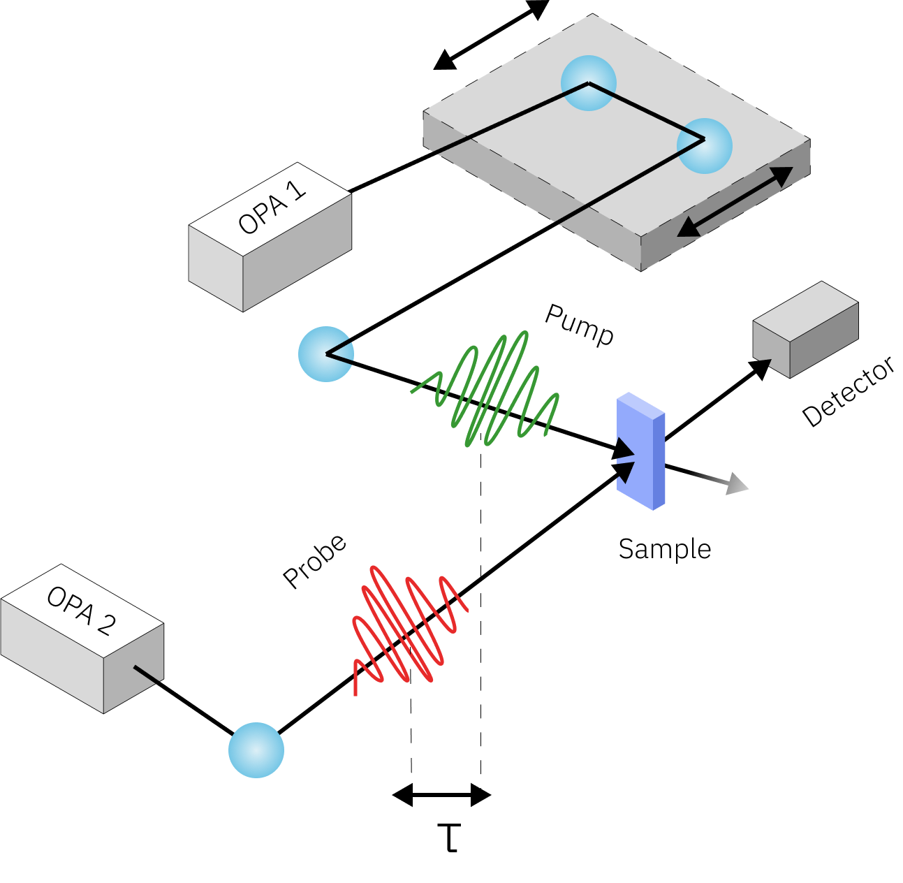
Different light sources can be used for these measurements: some examples include the use of a femtosecond light source whose beam is separated into two different paths for the pump and the probe. These types of setups implement Ti:sapphire lasers for excitation, with Optical Parametric Amplifiers (OPOs) for the selection of the emitted wavelength [Figure 2]. Other setups include different pulsed lasers for the pump and the probe, having pump pulses in the nanosecond to femtosecond time scale whose emission matches the absorption properties of the studied material, and probe beams emitted by broadband light sources.
Furthermore, the amplitude of the pump beam is typically modulated by an acousto-optic or electro-optic modulator; whereas the probe beam may be used in combination with a monochromator device, to select the wavelength of interest. In addition, dichroic beamsplitters, to combine the two beams; and galvanometric mirrors, to scan the sample with the combined beams are also used.
On the other hand, the detection system includes another dichroic mirror to separate the signal generated from the probe beam and the signal generated by the pump beam and a photodetector. The obtained signal is finally amplified by a phase-sensitive lock-in amplifier (Figure 2).
Requirements of the Technique
For the measurement of non-linear optical interactions, it is essential to have excitation light sources of high power and precise timing (ultrashort pulses). In this scope, mode-locked lasers can generate high-energy pulses in nanosecond to femtosecond time scales. Moreover, there is a trade-off between pulse duration and spectral bandwidth explained as the time-bandwidth product, for which one needs to take into account the relationship between time resolution and selection of the excitation wavelengths [5]. For this reason, narrow bandwidth lasers with femtosecond pulses are preferred.
As described in previous studies [4], several laser systems that have been implemented for pump-probe techniques include Ti:Saphire lasers working in ranges between 800 and 1064 nm with pulse widths of approximately 200 to 300 fs and repetition rates in the MHz scale. More examples include 830 nm of the pump beam and 1050 nm of the probe beam [6] to study red blood cells.
Furthermore, research on photosynthesis [5] describes the use of two types of lasers for transient absorption spectroscopy. The first family of lasers comprises sources with high energy pulses and low repetition rates (in the kHz range); whereas the second family comprises sources with pulse energies between 0.5 to 10 nJ and repetition rates in the range of the MHz.
The suitability and the benefits of this spectroscopy technique explain the great variety of examples and research fields in which it has been implemented. It allows for label-free imaging of molecules and nanomaterials that are non-fluorescent in a non-invasive way and with 3-D spatial resolution thanks to the third-order nonlinearity that is achieved. Additionally, pump-probe spectroscopy has found great use of interest in the Art field, where the study of colour pigments becomes possible thanks to the analysis of the absorption spectrum.
With the great number of possibilities that Pump-probe spectroscopy offers, the necessity of the most appropriate laser source seems evident. Ti:Saphire lasers have become the option of choice, however, these laser sources are unstable and may not be accessible to the general public.
We propose two interesting alternatives for pump-probe spectroscopy. The first one works in the femtosecond regime and consists of our cost-effective Cyclone laser, which solves the stability issues. Cyclone laser is a broad bandwidth fiber laser with extremely short Femtosecond pulses, which makes it possible for the generation of the third-order non-linearities that take place in pump-probe spectroscopy. It reaches between 15 to 20 fs pulse duration with optical peak powers larger than 120 kW. Additionally, it emits in a range between 950 and 1150 nm, which allows for the excitation of both biological and material science species, with deep penetration depths.
The second option of choice, working on the picosecond regime, is our Iceblink supercontinuum laser. It covers the Visible and NIR spectrum with a bandwidth between 450 and 2300 nm. The Iceblink emits less than 10 picosecond pulses with a total power of more than 3W, allowing it to characterize multiple materials without damaging the samples. Furthermore, it has its own accessory called Boreal, which selects wavelengths of interest in the Visible range and a minimum resolution of 5 nm.
