Research about Non-invasive optical techniques that use diffuse optics principles and their application for biomedical monitoring has become a central topic of discussion in the last decades.
With the development of new technology in the photonics field, with more sophisticated light sources and detectors, supercontinuum lasers have grown as a powerful tool in biomedical diffuse techniques. Because of the broad spectrum of these light sources, they allow for multiple applications in the medical field such as Near-Infrared Spectroscopy (NIRS). In this scope, Tachtsidis et al, 2021 described and compared the use of different supercontinuum light sources in Biomedical Diffuse Optics.
NIRS as a main tool in medical diagnosis – Contribution of supercontinuum light sources to this technique
NIRS is the most commonly used technique in nowadays medical diagnosis. It uses light sources that emit within the transparency window where optical absorption by tissues is diminished, favouring light scattering phenomena, enhancing the transmission of light within the tissue and so probing major utilities in the measurement of oxygenation in different tissues such as brain and muscle. However, in its most widespread configuration, this technique uses continuous wave illumination and is not able to provide information about the absorption and scattering coefficients, and the dynamical scattering properties of the tissue are not considered.
Another main limitation of CW-NIRS is related to the fact that because of the use of only a few wavelengths for excitation, not all the chromophores of the sample are considered during the measurements, reducing the accuracy of the technique.
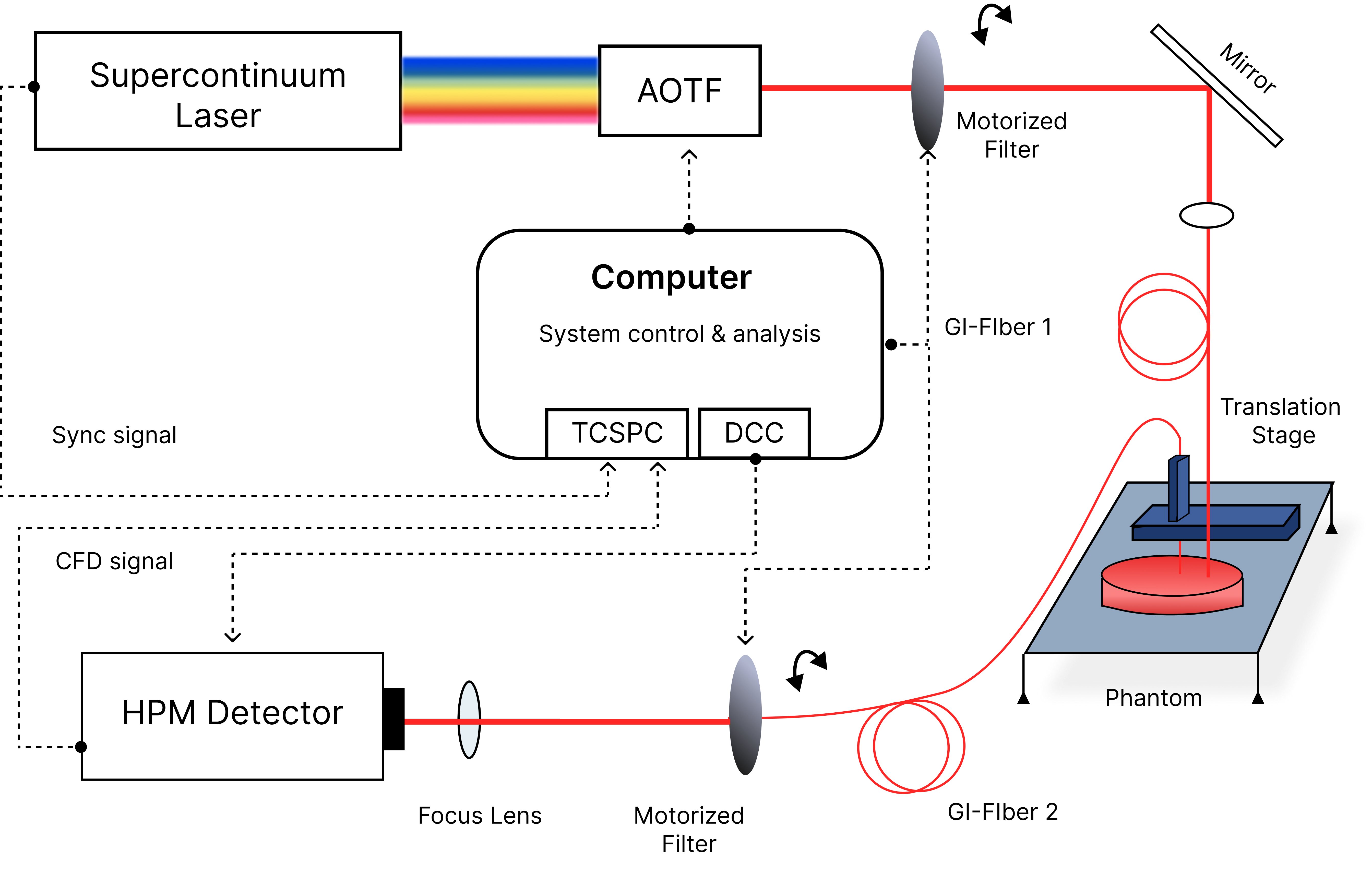
For this reason, Time Domain-NIRS is the most preferred approach, as it allows for discrimination of the information provided by different layers inside of the tissue. In TD-NIRS, a picosecond pulse reaching the sample is attenuated by the tissue and the re-emitted pulse is broadened, in the nanosecond scale. This technique provides a representation of the time-of-flight of the photons once they travelled through the tissue and were reflected back. From this information and the proper processing of data, accurate discrimination of the oxygenation levels from different tissue layers is achieved.
Although it is a well-established technique, the main challenge affecting TD-NIRS is the need for suitable equipment to be able to provide picosecond pulses, with enough power and fast detectors. Nevertheless, as described by Tachtsidis et al in their study, the development of supercontinuum lasers has made a major impact on the development of TD-NIRS. The reason for their study was related to the lack of standardisation in the NIRS technique, which requires a comparison of the available NIRS systems. They analyzed different Diffuse Optical setups and how research centres across the globe relied on the use of commercial supercontinuum light sources in medical diagnosis. An example of a TD-NIRS setup is shown in Figure 1 (courtesy of Lin Yang, Heidrun Wabnitz, Thomas Gladytz, Rainer Macdonald, and Dirk Grosenick, «Spatially-enhanced time-domain NIRS for accurate determination of tissue optical properties,» Opt. Express 27, 26415-26431 (2019)) One of the mentioned SCL was our Iceblink White light source.
Supercontinuum lasers´ features that are important for Biomedical Diffuse Optics
The Iceblink laser has become the option of choice in Biomedical Diffuse Optical techniques. The features that explain their potential include:
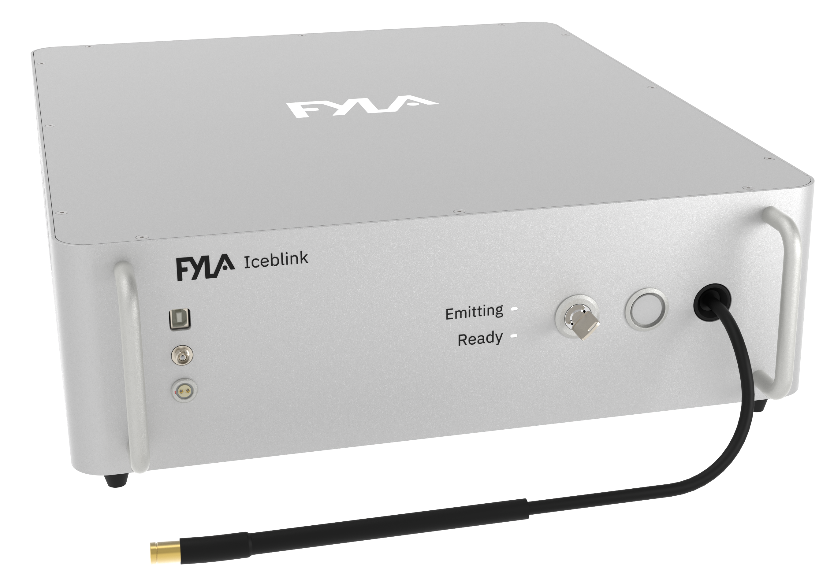
- Broad spectrum covering from 450 to 2300 nm. The spectrum of the Iceblink covers the entire VIS and NIR. This is an essential feature due to the fact that the Iceblink reduces the complexity and the costs of a setup. Because instead of using different narrowband sources, you can have all the wavelengths with just one laser. The result is a less complex and less bulky system and more cost-effective setups (see Figure 2 to get information about the dimensions of the Iceblink). Systems can be transported easily and become more practical for clinical applications. This advantage has led to the development of mammography systems and characterization measurements of different tissues such as bones and collagen, among others. There is also a potential for brain monitoring as one can perform real-time measurements of optical properties of the tissue over a large bandwidth and cerebral blood flow and perfusion measurements.
The pulsewidth of the Iceblink at FWHM is <10 picoseconds. It is within the range of pulse widths that have been reported as suitable for diffuse optical measurements. It allows for ultrafast dynamics measurements in tissues.
- The desired illumination system is the one having the narrowest bandwidth and the best spectral quality in terms of power stability and spectral density across the whole spectrum. In this scope, the Iceblink has its own accessory called Boreal, which enables the selection of wavelengths in a range between 450 and 750 nm. It i able to achieve a minimum bandwidth of 5 nm with excellent stability and no dispersion of the spectrum.
- The Iceblink provides >3 W of total power and >150 mW of power in the VIS range. These values are within the range of the powers that are needed to excite the biological samples without damaging them but achieving proper penetration depths. This power has been shown to be suitable for the monitoring of glucose dynamics and collagen.
For a detailed description of the latest trends in Diffuse Optical Spectroscopy for non-invasive tissue monitoring, click here to access the publication (https://www.mdpi.com/2076-3417/11/10/4616)