Towards a better understanding of the interaction between the photons of light and blood tissues, non-invasive and continuous techniques have been the point of focus of researchers in the last decades. The need for optical tools that achieve higher penetration depths without altering the samples suggests the use of diffuse optics.
Two well-known phenomena resulting from such interaction are absorption and scattering. Absorption consists of the dissipation of the photon energy; whereas scattering refers to the light being dispersed inside the tissue in random directions by the cells. The photons emitted by the light source are attenuated inside the tissue and the photon path length is a direct indicator of the depth sensitivity. In this scope, Diffuse Optics arise as a quantitative approach to explain photon migration inside of the tissue [1].
Several common techniques to study blood tissues include the traditional Diffuse Optical Spectroscopy technique (DOS) and the relatively new, Diffuse Correlation Spectroscopy (DCS). In Diffuse Optical Spectroscopy, also known as NIR spectroscopy, the attenuation of the photons at a specific wavelength is used as an indicator of blood volume and oxygenation. Whereas in Diffuse Correlation Spectroscopy (DCS), coherent and continuous wave NIR sources are used to study the scattering produced by the blood cells that are flowing inside of the tissues, producing a speckle pattern that changes proportionally to the movement of the blood cells, thus giving information about the index of blood flow [2].
Although these techniques are still widely used, they cannot reach the gold standard represented by more invasive techniques such as MRI, the reason for which Time-Domain methods are becoming more popular, thanks to the access to more sophisticated illumination and detection systems. [2]
Time-Domain Diffuse Correlation Spectroscopy
Changing to the Time-Domain means talking about time-of-flight: how long it takes for the photon to travel back from the tissue, in other words, it gives an estimation of the path length inside of the sample.
Time-domain diffuse Correlation Spectroscopy arises as a significant tool in clinical practice, resulting in a combination of Diffuse Correlation Spectroscopy (DCS) and Time-resolved Reflectance Spectroscopy (TRS).
In this technique, pulsed NIR lasers are emitted, and the light that is reflected or scattered back by the tissue is detected by a single-photon sensitive detector [3]. Measuring the photon time between the source and the detector gives an estimation of the time of flight, which is then used to calculate the Time Point Spread Function (TPSF).
Simaei et Al showed a prototype of a TD-DCS system [3]. Figure 1 shows the different components of a TD-DCS system.
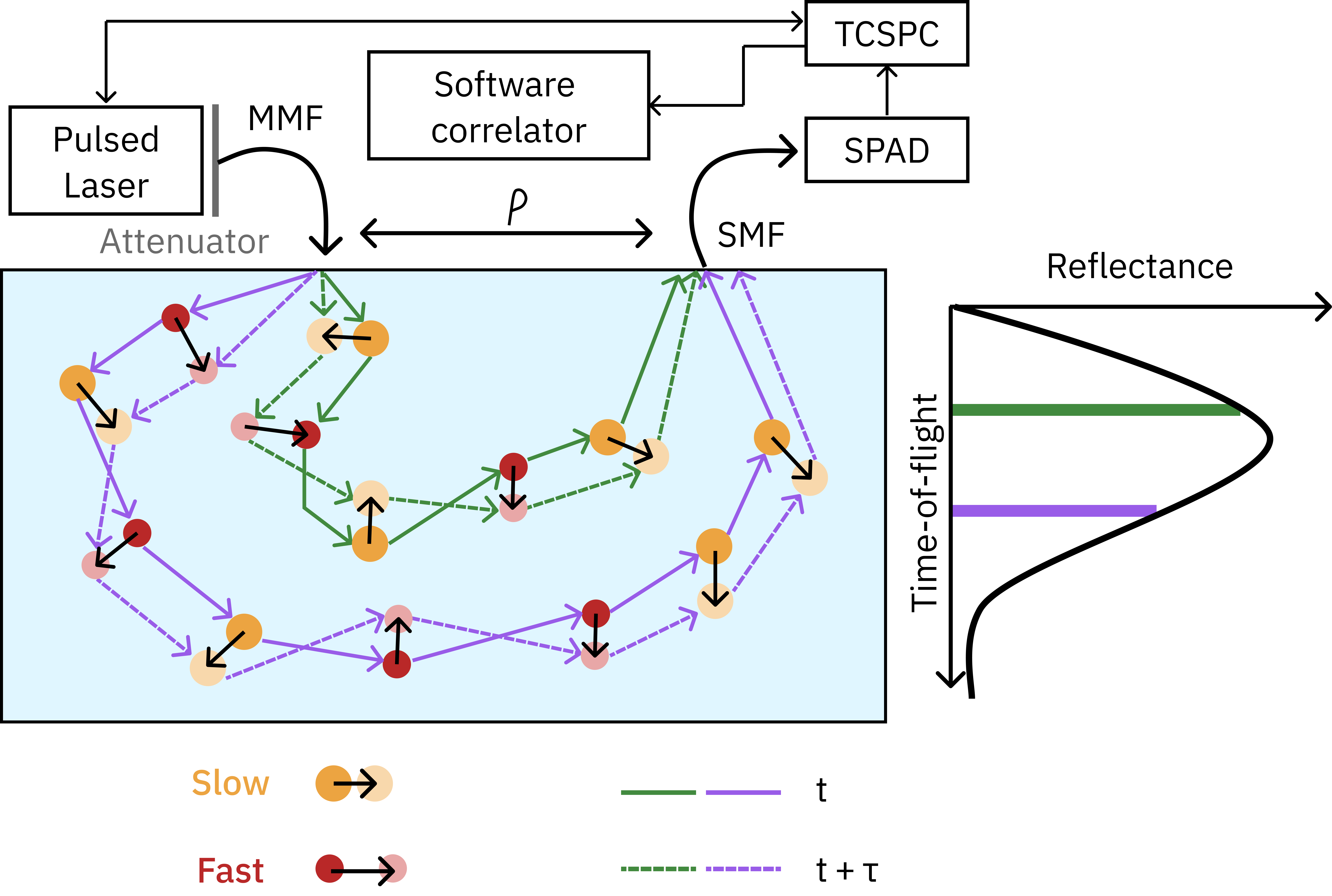
The main advantages of TD-DCS concerning CW-DCS include the possibility of not just studying the blood flow or optical properties but also acquiring more information about the dynamical changes and the Depth sensitivity that allows for obtaining more precise and selective information of deeper layers [1].
Important Instrument Considerations for TD-DCS
Three parameters determine the sensitivity and the Signal-to-Noise ratio of the measurements: the wavelength of the light source, the coherence length of the laser and the distance between source and detector. In this scope, several studies have been made in the comparison of the most suitable light source for such a technique [1,3, 5].
The first parameter is strictly related to the absorption and scattering inside of the tissue, having that longer wavelengths induce less absorption throughout the sample and thus, deeper penetration. Some studies suggest the use of emission at wavelengths between 750 and 780 nm [1,3]. Other recent experiments used emissions above the 980 nm of the water absorption peak [4]. Whereas others considered the use of tunable light sources emitting between 700 and 1030 nm [3].
The second parameter: coherence length, is limited by the pulsewidth of the laser, having that longer pulses induce longer coherence lengths. However, a compromise between coherence length and IRF (Instrument Response Function) needs to be considered. Shorter pulses are needed in order to improve the path length distribution of the photons at a certain time (path length distribution refers to photons that have travelled different paths throughout different layers of the sample). The physical limit of this compromise is given by the Time-Bandwidth product. It results in a balance between pulsewidth (and so, coherence length) and path length distribution (and consequently on the IRF).
Furthermore, the third parameter is the separation between the source and detector (also referred to as SDS). Traditional DCS or DOS requires an increase in the distance between the source and detector to obtain more information about the deeper layers of the sample and consequently reduce the number of detected photons. Time-domain techniques, on the other hand, allow for the discrimination between short and long path lengths. This results in SDS in the scale of tens of mm, compared to the standard 2-3 cm separation in DCS or DOS achieved with continuous wave (cw) lasers [6].
Some researchers have initially proposed the use of custom-built active mode-locked Ti:sapphire lasers emitting in the NIR obtaining interesting results [6]. Although the size of such light sources and the maintenance requirements, limit the use of the TD-DCS systems. On the other hand, other commercial pulsed lasers provide short coherence lengths, not being suitable for TD-DCS.
Conclusions show that optimal lasers would consist of picosecond pulsed lasers with long coherence lengths and narrow Instrument Response Functions.
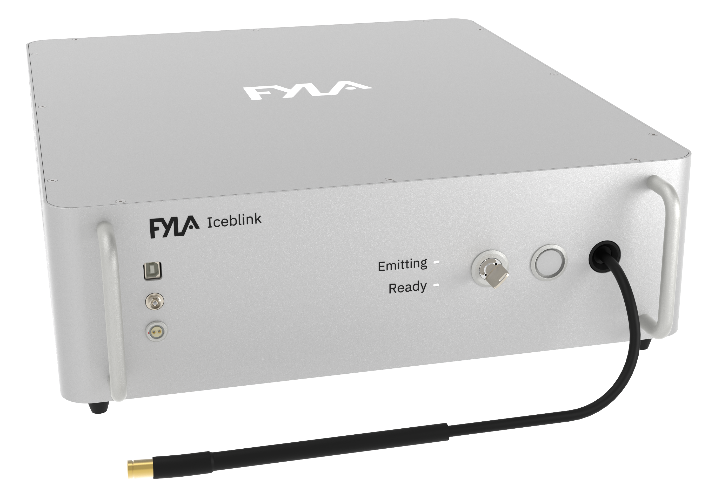
Iceblink Supercontinuum Laser Characteristics
In the search for the most convenient laser for TD-DCS, we propose the use of the Iceblink supercontinuum laser. The Iceblink supercontinuum laser has a broad spectrum, that covers the Visible and the NIR spectrum (450 to 2300 nm). With the appropriate filter set, one can select the desired wavelengths in the NIR, instead of having to use different monochromatic light sources for each experiment (ex: one laser for 750 nm, another laser for 780 nm, or another one for 1030 nm).
When it comes to the coherence length of the laser, not many studies have been provided regarding such parameters. Nevertheless, they are widely used in applications in which the coherence length is an essential parameter. The Iceblink supercontinuum laser has a total pulsewidth of 200 ps and a repetition rate of 80 MHz, which makes it a suitable tool in regard to the results obtained in the last publications [2, 3, 4, 6 ]. We estimate a theoretical coherence length of 40 mm.
The Iceblink supercontinuum laser does not require any maintenance and is a “plug-and-play” technology. It has an air-cooling system that would avoid the complex issues that arise when using Ti:sapphire lasers. In addition, its dimensions make it possible to optimize the size of your setup.