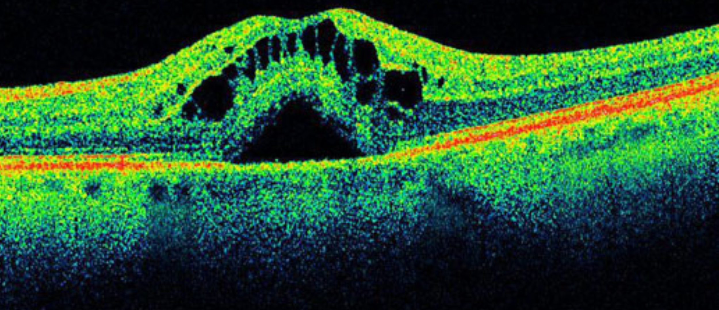
Fluorescence microscopy is a powerful imaging technique that allows you to visualize and study biological samples at the cellular and molecular levels. The technique relies on the use of fluorescent probes, which are molecules that emit light when excited by a specific wavelength. These probes can be attached to specific molecules or structures within a cell, allowing them to be selectively visualized and studied.
The enhancement of fluorescence image quality through meticulous slide preparation entails the adoption of numerous common steps, such as high numerical aperture lenses, low background staining protocols, or high-quality glass. Another critical component in this preparation process is the mounting medium, which assumes a pivotal role within this technique.
The mounting medium is a chemical substance that is used to seal the sample between the slide and the coverslip, preventing the sample from drying out and preserving its structure. It must be transparent, colourless, compatible with fluorescence probes, and with a precisely aligned refractive index.
Valeria Piazza et al., from “Centro de Investigaciones en Óptica” and “Universidad de Guanajuato” in México, show us in their paper “3,3′-thiodipropanol as a versatile refractive index-matching mounting medium for fluorescence microscopy” the importance of the refractive index property for choosing the right mounting medium and how they use Iceblink Supercontinuum for its obtention.
Exploring Refractive Index and Dispersion Curve with Iceblink Supercontinuum
The refractive index is a measure of how much a material bends light as it passes through it. Regarding the mounting media, the refractive index is critical because it affects the resolution and contrast of the microscope by minimizing optical aberrations.
A relevant facet to consider is that the refractive index of a material varies with the wavelength, and this variation is called dispersion. Because of this dependence, they considered it critical to select one light source which could measure in different wavelengths of the spectrum: FYLA’s Iceblink Supercontinuum.
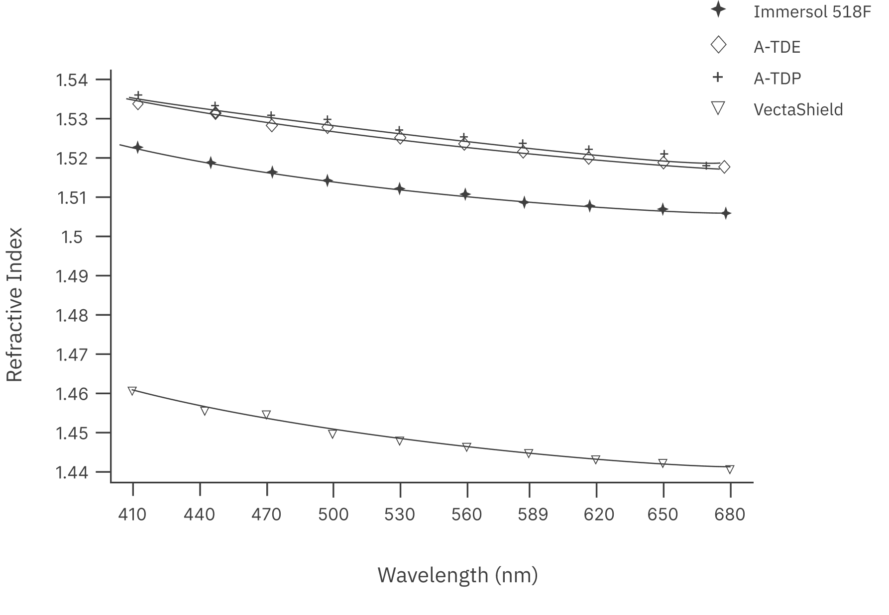
The primary objective was to determine the dispersion curve of different materials to study their different refractive index. The dispersion curve is a mathematical function that describes how the refractive index of a material changes with respect to the wavelength. The curve can be approximated using a mathematical equation, such as Cauchy’s dispersion equation, as illustrated in Figure 1.
Indeed, Iceblink occupies a pivotal role in this pursuit. FYLA’s Supercontinuum laser covers a broad spectral range, which is useful for measuring the refractive index dispersion curve of the materials over a wide range of wavelengths.
This specific study, (using a previous version of the Iceblink), allowed the creation of the dispersion curve measuring seven data points in the range of 500-680 nm in increments of 30 nm. The 590 nm point was replaced with 589 nm since it approximates the sodium line conventionally used for refractive Index reporting.
General Advantages of Iceblink Supercontinuum in Fluorescence Microscopy and Related Applications
The White Light Laser Iceblink is characterized by its spectral range from 450nm to 2300nm, covering both the visible and near-infrared regions. This feature is of immense value in fluorescence microscopy due to its exceptional versatility. Unlike Single-Wavelength lasers, the Iceblink’s ability to cover different excitation wavelengths allows the optimum selection of fluorophores with different absorption and emission properties and allows it to be used in a wide range of applications.
Briefly, this versatility has several advantages:
- Fluorescent Marker Selection: Iceblink makes it possible to excite a variety of fluorophores, each with a unique emission spectrum. This is critical for studying multiple components within a biological sample and for advanced research in cellular and molecular biology.
- Specific Labelling: By selecting the most appropriate fluorophore for the sample and experiment, specific and precise labelling of cellular structures and components is achieved. This improves the quality and accuracy of the resulting images.
- Application Flexibility: The broad spectral range of Iceblink provides flexibility for a variety of applications. It’s easier to see how Iceblink can be used for Dispersion Measurements and that could be employed as a light source for the Fluorescence Microscopy itself.
In addition to its versatile spectral range, Iceblink has the following features that contribute to its effectiveness in fluorescence microscopy and related applications.
- Total Power: With an output of > 3W, the laser generates sufficient intensity for strong and detectable fluorescence signals.
- Visible Power: With > 150 mW of visible power, the laser optimally excites fluorophores that emit in this critical region.
- Pulse Width: The laser emits light pulses of < 10 ps, enabling high-resolution temporal imaging of dynamic cellular events.
- Power Stability: Its exceptional stability, with variations of < 0.5% (std. dev.), ensures consistent and reproducible measurements.
The combination of these features makes the Iceblink laser an indispensable tool for adding to your setup.