Optical characterization with supercontinuum lasers allows for the measurement of fundamental parameters of different materials, which is a main requirement for the development and the correct performance of optical devices.
They make use of the property of the materials which consists of the fact that each molecule has its own absorption and emission lines, which depend on the electronic structure of a material. This means that for a certain molecule to be excited or to emit light (i.e. to undergo a transition), incoming light with a specific energy and wavelength is needed. This energy needs to match the energetic difference between the excited and the lower state inside of the atoms.
Microscopy techniques for optical characterization of devices
Some techniques that allow for the optical characterization of devices involve the use of microscopes. There are several types of microscopes, which can be classified according to the way in which the light reaches the sample. Consequently, some microscopes will operate using wide-field radiation, whereas others will scan the surface of the sample by means of a directed beam of light (i.e. light sheet microscopy). Additionally, other configurations include the use of scanning probes microscopy to analyse the surface of interest (i.e. Atomic Force microscopy or Scanning Tunnelling Microscopy). In the characterization of devices with microscopes, after the irradiated beam passes through the sample, the absorbed or emitted light is collected by the detection system of the microscope and optical images are generated.
NSOM technique for optical characterization
One emerging field that comprises an interesting scanning probe configuration is the NSOM or Near-Field Scanning Optical Microscopy technique, which is also referred to as SNOM or Scanning Near-Optical Field Microscopy. It consists of a method that tries to overcome the Abbe diffraction limit by means of using a nanoscale fibre probe to confine the light in a small area allowing for topographic and optical imaging at the subwavelength scale. For this reason, NSOM has been shown to be a useful technique not only for biological purposes but also for the characterization of different materials like semiconductors [1].
In this type of microscopy, light is delivered or collected by a probe, that can have the structure of a cantilever or the structure of a fibre probe.
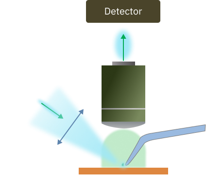
Additionally, the probe can operate in aperture or apertureless mode. In the apertureless mode, an AFM (Atomic Force Microscopy) probe that is coated with a metal to enhance the electromagnetic field at the part of the sample that is closer to its tip, is used in combination with an external light source placed at the far field for illumination (Figure 1a).
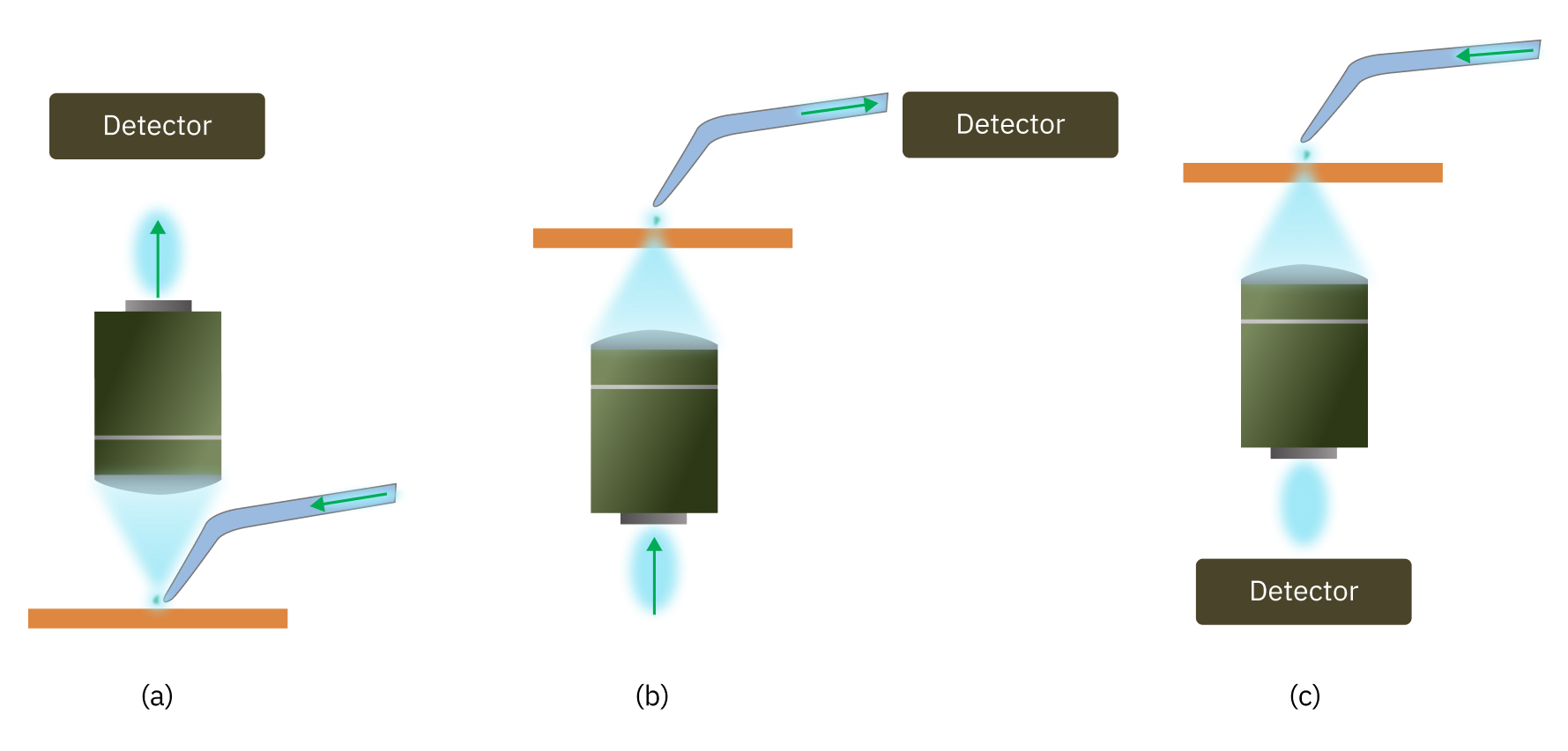
On the other hand, in the aperture mode, the light is delivered or collected through the scanning probe. There are different types of NSOM techniques that make use of aperture probes. The most simple methods include: Reflection NSOM (in which the light is delivered through the AFM probe from above the sample and the reflected light is collected) (Figure 2a), Collection NSOM (in which a light beam is focused on the sample from below and the probe is placed next to the surface, so that the transmitted light passes through it to reach the detector) (Figure 2b) or Transmission NSOM (in which the sample is irradiated through the probe from above and the light that is transmitted through it is collected) (Figure 2c).
Requirements for the NSOM technique
Regarding the improvement of the resolution of an NSOM system, it is necessary that the illumination source is focused down towards the excitation spot of the sample in a stable and efficient form. This is the reason why coherent illumination sources are preferred over other possibilities. Several light sources that have been used traditionally include emission ranges between UV and IR wavelengths with optical powers between 0.5 and 5 mW, showing that IR wavelengths allow for better confinement of the light. Moreover, both CW and pulsed lasers have been implemented. Some examples include the use of Ti:Saphire lasers in combination with supercontinuum generators. Other examples implement more than one gas laser source at the same time, although recent studies have proposed ultrafast pulsed laser as an outstanding technology for SNOM measurements [2].
The reason for that resides in the generation of very short pulses that allow for the measurement of the dynamics of molecules in such short time scales.
In this scope, we present our alternative, called Iceblink. With the Iceblink it is possible to excite the surface at a wide range of wavelengths (from 450 to 2300 nm), with pulses that are shorter than 10 picoseconds in the central wavelength and combine it with a tuneable filter when a specific linewidth is needed. In this scope, the Iceblink could be used in combination with its accessory: the Boreal, which uses the bandpass technology to filter wavelengths with a minimum of 5 nm resolution. The system comprising the Iceblink and the Boreal is ideal for NSOM technology in life and material science due to the broad and flatten spectrum of the laser, which allows for different fluorophores or chemical compounds in a heterogeneous sample to be excited and imaged at the same time with great stability (<0.5 % std. dev); and the possibility of selecting wavelengths of interest with the Boreal accessory.